


HEBREW UNIVERSITY OF JERUSALEM · FACULTY OF MEDICINE

How organelles talk, adapt & survive.

We investigate how cells sense and adapt to mitochondrial and peroxisomal stress, how organelles communicate with one another, and how these pathways shape physiology, aging and disease.

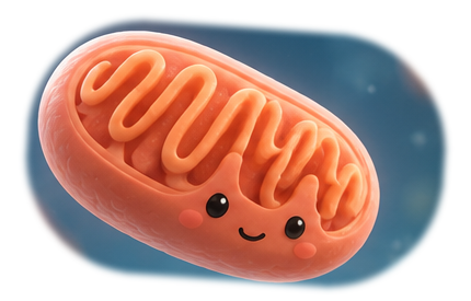
Illustrated mitochondrion
Illustrated peroxisome
mitochondrion ↔ peroxisome

Research Support

Our research is supported by:

Israel Science Foundation (ISF)
United States–Israel Binational Science Foundation (BSF)

RESEARCH

Stress responses across organelles.

Peroxisomes

Peroxisomal stress responses

Peroxisomes are vital organelles, involved in various metabolic processes. Despite their critical roles, our understanding of how cells respond to peroxisomal stress remains limited. Our primary goal is to address these gaps and shed light on the quality control pathways activated during peroxisome dysfunction. Through the development of temperature-sensitive in vivo worm models for peroxisome biogenesis disorders and tissue-specific degron-based knockdowns, we aim to regulate peroxisome function spatially and temporally. This approach provides a robust platform for a comprehensive exploration of nematode responses during peroxisome dysfunction and recovery. Additionally, we utilize mammalian cell culture techniques and cells obtained from patients with peroxisome biogenesis disorders to conduct a thorough characterization of peroxisome stress responses.

Diagram illustrating peroxisome stress responses and transcriptional response to the nucleus
Mitochondria

Mitochondrial stress responses

Mitochondria are essential double membrane organelles, involved in nearly every aspect of our lives including energy production. Dysfunction of this organelle is associated with a vast array of clinical manifestations including neurodegenerative and metabolic disorders, the aging process and muscle loss. There is no cure for mitochondria disorders. Our goal is to characterize how cells cope with mitochondria dysfunction, what are the stress responses that are activated during mitochondrial stress and whether unregulated activation of these responses contributes to our health or to the development of diseases.

Fluorescence microscopy image illustrating mitochondrial stress responses
Aging
Cancer
Neurodegeneration
Development
Communication
Biogenesis
Maturation

PEOPLE

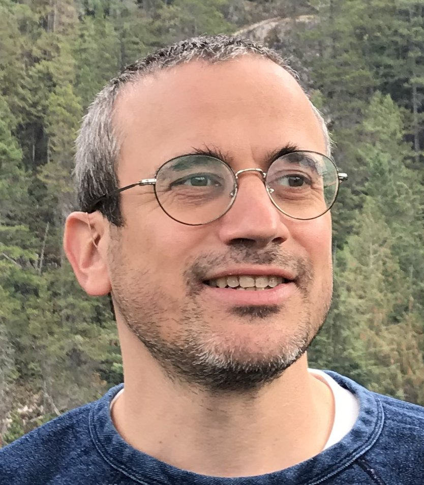
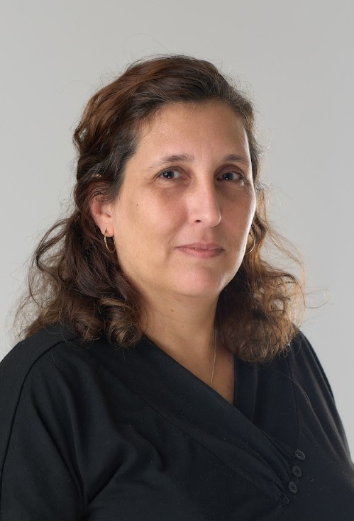
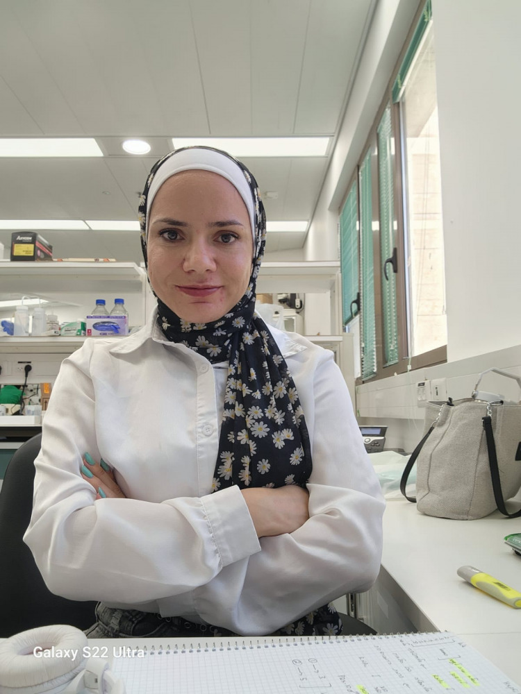
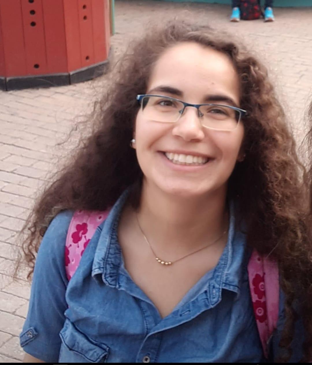
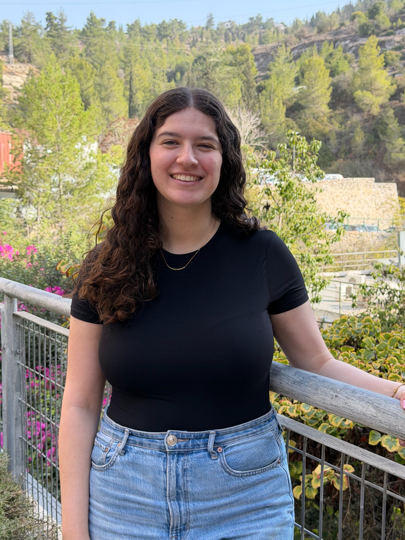
The Shpilka Lab team.

A small group with a shared interest in organelles, stress biology and experiments that connect molecular mechanisms to physiology.

LAB LIFE

Beyond the bench.

A glimpse of life in the Shpilka Lab — experiments, conferences, trips, celebrations and everything in between.

2023

The homodimer interfaces of costimulatory receptors B7 and CD28 control their engagement and pro-inflammatory signaling

Andrey Popugailo, Ziv Rotfogel, Michal Levy, … Tomer Shpilka, Raymond Kaempfer

Journal of Biomedical Science · 30 · 49

2022

ATFS-1 regulates peroxisome assembly genes and protects both mitochondria and peroxisomes during peroxin perturbations

Tomer Shpilka, Nandhitha Uma Naresh, Yunguang Du, Jennifer L. Watts, Cole M. Haynes

bioRxiv

LONP-1 and ATFS-1 sustain deleterious heteroplasmy by promoting mtDNA replication in dysfunctional mitochondria

Qiyuan Yang, Pengpeng Liu, Nadine S. Anderson, Tomer Shpilka, et al.

Nature Cell Biology · 24 · 181–193

Mitochondrial genome recovery by ATFS-1 is essential for development after starvation

Nandhitha Uma Naresh, Sookyung Kim, Tomer Shpilka, Qiyuan Yang, Yunguang Du, Cole M. Haynes

Cell Reports · 41

2021

UPRmt scales mitochondrial network expansion with protein synthesis via mitochondrial import in Caenorhabditis elegans

Tomer Shpilka, Yunguang Du, Qiyuan Yang, Andrew Melber, et al.

Nature Communications · 12 · 479

2020

Histone deacetylases 1 and 2 silence cryptic transcription to promote mitochondrial function during cardiogenesis

Zachary J. Milstone, Sherin Saheera, Lauren M. Bourke, Tomer Shpilka, et al.

Science Advances · 6 · eaax5150

2018

The mitochondrial UPR: mechanisms, physiological functions and implications in ageing

Tomer Shpilka, Cole M. Haynes

Nature Reviews Molecular Cell Biology · 19 · 109–120

2015

Fatty acid synthase is preferentially degraded by autophagy upon nitrogen starvation in yeast

Tomer Shpilka, Evelyn Welter, Noam Borovsky, Nira Amar, et al.

PNAS · 112 · 1434–1439

Lipid droplets and their component triglycerides and steryl esters regulate autophagosome biogenesis

Tomer Shpilka, Evelyn Welter, Noam Borovsky, Nira Amar, et al.

The EMBO Journal · 34 · 2117–2131

Lipid droplets regulate autophagosome biogenesis

Tomer Shpilka, Zvulun Elazar

Autophagy · 11 · 2130–2131

2012

Essential role for the mammalian ATG8 isoform LC3C in xenophagy

Tomer Shpilka, Zvulun Elazar

Molecular Cell · 48 · 325–326

Mechanisms of autophagosome biogenesis

David C. Rubinsztein, Tomer Shpilka, Zvulun Elazar

Current Biology · 22 · R29–R34

Ubiquitin-like proteins and autophagy at a glance

Tomer Shpilka, Noboru Mizushima, Zvulun Elazar

Journal of Cell Science · 125 · 2343–2348

2011

Atg8: an autophagy-related ubiquitin-like protein family

Tomer Shpilka, Hilla Weidberg, Shmuel Pietrokovski, Zvulun Elazar

Genome Biology · 12 · 1–11

Binding of superantigen toxins into the CD28 homodimer interface is essential for induction of cytokine genes that mediate lethal shock

Gila Arad, Revital Levy, Iris Nasie, … Tomer Shpilka, Raymond Kaempfer

PLoS Biology · 9 · e1001149

LC3 and GATE-16 N termini mediate membrane fusion processes required for autophagosome biogenesis

Hilla Weidberg, Tomer Shpilka, Elena Shvets, Adi Abada, et al.

Developmental Cell · 20 · 444–454

Shedding light on mammalian microautophagy

Tomer Shpilka, Zvulun Elazar

Developmental Cell · 20 · 1–2

2010

LC3 and GATE-16/GABARAP subfamilies are both essential yet act differently in autophagosome biogenesis

Hilla Weidberg, Elena Shvets, Tomer Shpilka, Frida Shimron, et al.

The EMBO Journal · 29 · 1792–1802

Mammalian Atg8s: one is simply not enough

Hilla Weidberg, Tomer Shpilka, Elena Shvets, Zvulun Elazar

Autophagy · 6 · 808–809

JOIN US

Come ask organelle questions with us.

We welcome inquiries from motivated MSc and PhD students, postdoctoral researchers and other scientists interested in organelle biology, stress responses, C. elegans, mitochondria or peroxisomes.

Interested in joining?

Send a short introduction, your scientific interests and a CV.

Email the lab

CONTACT

Find us in Jerusalem.

Department of Biochemistry & Molecular Biology · Faculty of Medicine · Hebrew University of Jerusalem.

Shpilka Lab

Butner Building · First floor · Room 127
Hadassah Ein Kerem campus
Jerusalem 9112102 · Israel

Lab: +972 2 675 7348
Office: +972 2 675 8283

tomers@ekmd.huji.ac.il